Friday, February 17, 2017

Reduction in Ophistorchis numbers and Th1 responses: correlation or causation?


This post discusses the following paper, chosen by a classmate for her presentation in a class that was about the pathogenesis of persistent infections:
Kaewraemruaen C, Sermswan RW, Wongratanacheewin S. CpG oligodeoxynucleotides with crude parasite antigens reduce worm recovery in Opisthorchis viverrini infected hamsters. Acta Trop 2016;164:395-401


Opisthorchis viverrini is a food-borne liver trematode, found in South east Asia, giving it the common name - Southeast Asian liver fluke. It has a typical trematode life-cycle (See CDC's page here). Infection itself can be divided into the acute and chronic phases, with the acute phase having a typical Th1 type immune response, characterized by IL12 and IFNg, and the chronic phase having a typical Th2/Treg response with IL4, IL 10 and TGFbeta. Hamsters are used as models to study the immunopathology of this infection.

In a recent paper published from Thailand, Kaewraemruaen et al 1, in trying to answer the question of the role played by Th1 responses, hypothesize that there might be a protective role played by Th1 cytokines in the early stages of infection. The hypothesis is derived from the fact that in the acute phases of infection, Th1 related cytokines are upregulated, and that hamsters vaccinated against O. viverrini have shown a Th1 skewed response, with low amounts of Treg cytokines IL10 and TGFbeta. In the later stages of infection, the adult parasite skews the response to a Th2/Treg type, and persists in the body, causing the chronic infection.

The methodology used was to induce a Th1 response using an adjuvant that typically induces such a response and determine if the response caused a reduction in the number of adult trematodes recovered after a time period.

Evidence to prove their hypothesis came from the following experiments:
a.  In vitro proliferation of, and IFNg production from mononuclear cells from naïve hamster spleens were higher for the Th1 cytokine inducing adjuvant CpG Oligodeoxynucleotide than the non-CpG oligodeoxynucleotide control and the crude somatic antigen, but lower than the assay positive control (stimulation with PAM and ionomycin) 72 hrs after incubation.
b. Hamsters that had been injected with CpG ODN alone or with crude somatic antigen or somatic antigen alone, had higher numbers of CD4+ T cells, higher amounts of IFNg and higher numbers of IFNg+ T helpers, than the negative controls, although the presence or absence of somatic antigen with the adjuvant had no bearing on the numbers of CD4+ cells.
c. Despite the adjuvant only animals exhibiting robust Th1 responses, number of adult worms recovered from the hamsters was reduced only when the adjuvant was combined with the crude somatic antigen, and this reduction was not statistically significant. However, there were fewer adult worms in animals that received that adjuvant and adjuvant + antigen compared to PBS injected control animals.

The paper is also significant in that the authors used mAbs directed against murine IFNg to cross detect IFNg in hamsters (both IFNg proteins have 56% identity in their primary structures). Also, CpG ODN which acts through TLR 9 in mice to stimulate innate immune cells was able to act on TLR 9 of hamsters, which is expected given the idea of conservation of TLRs across phyla, and the fact that both animals are rodents, and hence closely related.

Thus, the authors sort of show that a Th1 response is protective, as it apparently is in Schistosoma mansoni infections. However, we get into the semantics of what "protection" actually means. My expectation at the beginning of my reading was that the authors meant protection against reinfection, since the Th1 clearly was an acute phase response. For reasons unclear, the conclusion is that a Th1 response causes protection in the acute phase of the disease, with "protection"being defined as a reduction in the number of adult parasites. However from a disease perspective, no histopathology was done to show that the insult to the liver was any different because of the response. Also, it was not shown in any way that the increase in IFNg+ T helpers were specific to the trematodes and were not just randomly activated T helpers found in blood that had little to no impact on the adult trematodes in the liver. The role of other non- Th1 based responses must have also been considered, to explain the reduction in adult parasite numbers.

Kaewraemruaen C, Sermswan RW, Wongratanacheewin S. CpG oligodeoxynucleotides with crude parasite antigens reduce worm recovery in Opisthorchis viverrini infected hamsters. Acta Trop 2016;164:395-401.


Tuesday, March 8, 2016

"You need all sorts" - Lessons from the deer tick genome


A wise person in Fargo, ND, once told me, “You need all sorts”. The adage seems to be true in parasitology. You need all sorts of studies to understand parasitosis. Epidemiological studies that assess risk factors, microepidemiological studies that describe new haplotypes, drug studies of anti-parasitological agents, studies on parasite behavior and biology, novel techniques to diagnose and control parasites, new applications of old techniques – all of these are needed, and more. Among the latter are extensive studies that map out entire genomes of parasites. As always, the early scientist gets the worm, or in this case, the acarine.
  
The genome of Ixodes scapularis, the deer tick, one of the most feared ticks in North America, has been sequenced and insights were revealed to the general public in a recent paper published in Nature Communications, entitled ‘Genomic insights into the Ixodes scapularis tick vector of Lyme disease’ by an impressive knot of ~93 authors, the who’s who of tick biologists on the planet. 

File:Ixodes.scapularis.jpg 
(Source of image: Sandy Rae, Wikimedia Commons. This file is licensed under the Creative Commons Attribution-Share Alike 3.0 Unported License)

The key features of interest from the publication have been summarized below.
  
Large haploid genomes seem to be the norm, and by “large”, the authors mean >1Gbp. That is 1, followed by 9 zeros. Previous studies by Ullmann et al in 2005, estimated the genome size to be around 2.1 x 10^3 Mbp, with 34% of the DNA being unique. The other part had been found to be made up of highly repetitive (27%) and moderately repetitive elements (39%). A summary of the assembly statictics in the Nature Communications paper estimates that the I.scapularis genome has 20,486 genes, with a mean length of 10,589 bp, and an average coding sequence length of 855 bp. 60% of the genome has orthologous genes in other species, with 22% of the genes being paralogs.

The authors used FISH based karyotyping to identify 26 acrocentric autosomes and 2 sex chromosomes. Their experiments also revealed a high percentage of tandem repeats in the centromeres and pericentromeric areas, which is in line with the previously described repetitive areas. To look at some beautiful karyograms, head over to the original paper.

Class I and II transposable elements were also found. Annotators found 41 long terminal repeat reterotransposon family elements, 37 Ty3/gypsy group, 234 miniature inverted-repeat transposable elements, Mag, CsRn1, Squirrel, Toxo, mariner and piggybac.

So, why should you and I care? What great insights does the genome reveal? What lessons can a fellow parasitologist learn?
  
Lesson 1: Once bitten, twice shy
When ticks feed, they inject saliva into the feeding area. And tick saliva is more complex than that of mosquitoes. The authors believe that 0.4% of the predicted proteome of I.scapularis, that is, about 74 proteins, contain a Kunitz domain. Kunitz domain-containing proteins essentially are protease inhibitors, and so play roles in inhibiting coagulation. Since even mosquitoes have less than 10 of these protease inhibitor proteins, it is easy to believe how effective the tick saliva is in inhibiting coagulation, angiogenesis and vasodilation.  The authors also found about 40 lipocalin genes in the saliva, that have anti-inflammatory properties, and 34 metalloprotease genes, all ensuring that the blood, like wine, keeps flowing at the tick banquet.

Lesson 2: Blood is thicker than water, till it is lysed
It is well known that ticks ingest blood. What is not so well known is that RBCs hemolysed in the tick midgut are pinocytosed into gut epithelial cells, where enzymes like cathepsin D, cathepsin L and serine carboxypeptidase aid in the digestion of haemoglobin, releasing heme, which is curiously spelt as “haem” in the article, which is transported to “haemosomes” to be detoxified and stored as hematin aggregates. The authors were able to identify the genes that code for the enzymes that orchestrate the above.

Lesson 3 : “I can’t make it on my own, I need you”, says Ixodes scapularis
The authors identified genes that coded for two families of haem storage proteins, since ticks are unable to synthesize haem de novo. These two families – haemlipoglyco-carrier proteins and vitellogenins protect the tick from the dangers of a haem rich diet, and help store haem for the embryo. Essentially, the iron that the tick embryo  needs for haem synthesis is supplied by the host indirectly.

Lesson 4 : I’m not crazy, my reality is just different than yours
Neuropeptides act via their corresponding GPCRs. From the genome of I.scapularis, the authors identified 39 neuropeptide genes, which play important roles in molting, synthesis and hardening of the cuticle, diuresis, development and reproduction, and to a large extent behavior and life styles. So, why in the world is this important, you ask ? Well, the 100 fold size increase in size of the tick body after feeding, must be accommodated, and these molecules – buriscon alpha and beta, corazonin and eclosion , are vital. The G protein coupled receptors that these neuropeptides associate with are also numerous. The authors were able to identify GPCRs like kinins, inotocin, tachykinin, allostatin-A etc. Given the different essential conditions needed to thrive in a tick world, it makes sense that the crazy numbers and amounts of neuropeptides synthesized by the ticks, contributes to a complexity that is not seen in many higher taxa.

Lesson 5 : Growing pains
To start vitellogenesis, ticks use molecules called ecdysteroids. These have names like “disembodied”, “Halloween” and “spook”/”spookier”. Genes for enzymes of the mevalonate-farnesal and other pathways involved in the production and reactions of juvenile hormone III were also found to be present.

Lesson 6: You purpose to kill me. How dare you sport thus with life?
The authors found a record number of detoxification genes of the CYP450 fame (206 genes) and a whole lot of carboxylesterase/cholinesterase-like genes (~75 genes). They postulate that ticks may need to detoxify toxic factors in the blood of hosts that they feed on and also substances that they encounter off the host. But those numbers seem incredible when compared to the body louse, which has 36 genes with detoxification functions, but also feeds on blood.

Lesson 7: Grandma, what a large hypostome you have! All the better to taste you with, my dear!
The rule is, ticks have to first find their hosts before they can feed on their (host) blood. They set out on their quest equipped with an impressive array of sensory organs. In the genome, 62 gustatory receptor genes, and 29 ionotrophic Glutamate receptors were found. Cuticular lipids, and non-volatile mounting pheromones in the females and males are suspected to be produced, that aid not just in questing for hosts, but also another member of the species.

Lesson 8: The eye is the lamp of the body
As far as eyes go, ticks don’t have large complex eyes like humans, but only have “photon- sensitive receptors”. Opsin G protein coupled receptors were identified in the genome, and are thought to be involved in long-wavelength light perception. The authors could not identify UV and short wavelength receptors, postulating that since ticks do not rely on vison to locate hosts, mates and oviposition sites, the sensations accorded by the highly developed thermal, mechanical, and olfactory receptors were enough.

Lesson 9: Of parasites, vectors and defense against the dark ones
As vectors extraordinaire, ticks are capable of carrying pathogens. But, it would be highly beneficial for them to not be afflicted by these same pathogens that they so willingly ferry from one host to the next susceptible one. The genome encodes for the appropriate orthologs of Toll, Jak-STAT, AMPs, caspases, akirins, ixoderins, lysozyme and RNAi genes (especially Ago genes, but upto 121 unique genes that may be involved in anti-viral defense), among others, all of which protect the tick from the pathogens of vertebrates.

The ticks also apparently take good care of the pathogens that they transmit, by synthesizing proteins and receptors that aid in their survival and transmission, such as Salp15, Salp20, receptor for Borrelia lipoprotein BBE31, P11, among others. Pathogens take full advantage of the hospitality offered by helping the tick, or by being a disruptive visitor. Anaplasma helps the tick survive better in cold climates, by upregulating putative “anti-freeze” genes. But being dual faced, it also inhibits apoptosis in the tick gut, establishing infections in them, and using other mechanisms, such as protein misfolding to evade cellular responses.

When a pathogen is a bad guest, tick responses include, but are not limited to, extrinsic apoptosis pathway induction in the salivary glands, decreasing glucose metabolism, changing the way protein is processed, producing HSPs, and increasing subolesin.

Lesson 10: Never compare, unless you absolutely have to
The authors used quantitative proteomics to identify differences between tick-Anaplasma interactions in actual infections, and in an artificial infection in a tick cell line. They found that at least 83 proteins were different, with differential gene expression being evident over the course of the infection. (Please look at the supplementary materials in the original paper for details)

Wrapping up with some epidemiology
The authors also studied the population structure of I. scapularis in North America, by studying SNPs from 8 populations, from the USA. Northern and Southern populations were found to be considerably different, compared to the mid- west (WI and IN) populations. There was also differences between the lab adapted Wikel strain and wild types from the field.

So, what now? What next?
The authors were able to identify novel genes that are potential targets for acaricidal drugs, developing drugs against which would ensure the selective toxicity we desperately seek with chemotherapeutics. The paper is a illuminating in the sense that it is not a list of genes. Genomic finds have been beautifully tied to function. With ~93 authors, 38 supplementary tables, 25 supplementary figures, and no doubt hours of mental and CPU labor, we don’t expect any less.
 
References:
M. Gulia-Nuss et al., Genomic insights into the Ixodes scapularis tick vector of Lyme disease. Nat Commun 7, 10507 (2016).
A. J. Ullmann, C. M. Lima, F. D. Guerrero, J. Piesman, W. C. Black, Genome size and organization in the blacklegged tick, Ixodes scapularis and the Southern cattle tick, Boophilus microplus. Insect Mol Biol 14, 217-222 (2005).

Saturday, November 14, 2015

Tapeworm tales and lessons in public health

This post is for my friends and family who are not veterinarians.

Tapeworm news periodically make the rounds. I remember asking my mother when I was 14 (and suffering from a bout of headaches), "What if I have a tapeworm in my brain like Leander Paes?" A MRI at a big Bangalore hospital revealed that I didn't. But, Paes, a Grand Slam-winning, star doubles-tennis player, who had contracted it in the Unites Sates, did. And it had been all over the news (News story here).

More recently, a 26- year old man living in California's Napa valley, was diagnosed with the condition "neurocysticercosis" (caused by the tapeworm Taenia solium), when he went to the emergency room complaining of a headache. Following his surgery and subsequent recovery, the man told the news channel this : “I just couldn’t believe something like that would happen to me. I didn’t know there was a parasite in my head trying to ruin my life.” (News story here). Whether the parasite was "trying to ruin" his life is a question that no one can answer. It was doing what it was wont to do, after all.The quick and brilliant diagnosis and treatment given to the man by the ER doctors saved the man's life.

Other recent stories have included the case of dwarf human tapeworm adults, Hymenolepis nana, in an immunocompromised human adult,  that had showed malignant transformation (the worm had the tumor, that is). Here is the original research article in the New England Journal of Medicine (link here). And all at once, the  "experts" who are neither veterinarians nor human physicians, have jumped on it, having a field day. The word "immunocompromised" seems to have been left out in news reports, and a whole host of blogposts have been written by these self professed "health writers", who have obviously done a lot of research on that veritable fount of wisdom and knowledge, called Google. The results speak for themselves, and the sole purpose appears to be fear mongering (like this one here).

In popular culture, neurocysticercosis has been featured in an episode of the popular show "House". Even comics and memes feature these dorso-ventrally flattened creatures (Comic here). https://upload.wikimedia.org/wikipedia/commons/e/e2/Taenia_saginata_adult_5260_lores.jpg
"Taenia saginata adult 5260 lores" by http://phil.cdc.gov/PHIL_Images/20031208/87d4bff74e41427cb278526bd9cbe76a/5260_lores.jpg. Licensed under Public Domain via Commons - https://commons.wikimedia.org/wiki/File:Taenia_saginata_adult_5260_lores.jpg#/media/File:Taenia_saginata_adult_5260_lores.jpg
  
As a Veterinarian and as a parasitologist, who does actual research in the field, I recommend a sane, balanced evaluation of  tertiary literature, especially of blogposts and newspaper articles. In other words, don't believe everything written by self proclaimed "health experts", who are only arm-chair philosophers, who got their information from the first ten search results that Google (or worse Bing) brought up. [The letters that come after a person's name, gained after years of structured instruction at an academic institution, actually mean something. And if those letters are not MD, DVM, BVSc or something equivalent, it is best not to take parasitology advice from the people whose names precede the letters. (I assure you that we are not part of a conspiracy to get you)]

So, here are some things that you might be wondering about, which I had taken the liberty of answering, before you have voiced your questions.

*Q: Do humans get tapeworms?
 A: Why, yes! They do. Both the tapeworms mentioned above (Taenia solium and Hymenolepis nana) are human tapeworms.

*Q: Are the two tapeworms mentioned above the only ones that humans get?
A: No, humans can be infected by other tapeworms too. If you ingest dog fleas that have the larval form of the dog tapeworm Dipylidium caninum, you can get infected by the adult of that species. If you eat uncooked fish with the larval stages of Diphylobothrium latum, you can be infected by adults of that species. If you drink water that have Cyclops or live microscopic floatsam that have the larval stages of Spirometra mansonoides , you can become the intermediate host for that tapeworm. If you ingest dog feces accidentally (I hope no one ingests dog feces intentionally), and if there were Taenia or Echinococcocus eggs in that feces, you can become the intermediate host for those tapeworms. There are other tapeworms that humans could potentially get, but notice how the words uncooked and feces keep recurring.
 
*Q: Practically speaking, did the man with the tapeworm cancer, actually have cancer?
A: No, the tapeworm had the cancer. Let me paraphrase that. The tapeworm did not cause a cancerous growth. It had the cancer itself. The man had HIV, and so his immune system could not do anything to prevent the spread of the cancerous tapeworm cells into his lymph nodes.

*Q: Are there many other humans who have tapeworm cancers that have been misidentified as human cancer?
A: Probably not. The case report in NEJM is the first of its kind reported. It began when pathologists who were looking at lymph node biopsies from the man realized that the neoplastic cells were smaller than human cells. Since pathologists are trained to look for such things, and since staging cancers involves these trained clinical pathologists to deliver their verdict on the malignancy of the cancer before the institution of treatment, you can be assured that they will be able to identify such things.

*Q: Can you/I get tapeworms that get cancers?
 A: Not if you are a healthy adult. Generally, adult tapeworms cause very little effect on the their adult hosts. Unless you have hundreds of them, you will probably not even know that you have them.

The only way to get hundreds of them is to eat hundreds of the infective tapeworm larvae present in pork muscle. See the little white spots in the picture below? Those are tapeworm larvae. Know and recognize them.
 
(Source : http://www.austincc.edu/microbio/2704q/ts.htm)

Or in the case of Hymenolepis nana, eating the eggs shed by another human in his/her stools, who harbours the adult tapeworms in their intestines (who has not washed their hands after defecation).


*Q: If you have in the recent past eaten uncooked pork, how do you know if you are have tapeworms in your intestines?
A: Tapeworms live in the intestine and either shed eggs or shed their segments that burst open to release eggs. These get into the environment through the feces of the host. Feces can be examined under a microscope to see if you have tapeworms inside.

*Q: Are there treatments to get rid of adult tapeworms?
Yes. There is very effective medication approved for human use and for animal use, that can be prescribed to infected patients.

*Q: If prevention is better than cure, how can neurocysticercosis, and infection with adult tapeworms be prevented?
Here are some ways to help you avoid tapeworm infections:
1. Dispose human and pet animal waste properly.
2. Make sure that meat of all kinds is cooked properly. If you find that your beef or pork is "measly", do yourself a favor and throw it out. Make sure no one else eats it either.
Rare steaks can result in rare cases of tapeworms.
3. Alternately, freeze meat at -10C or less for more than 48 hrs.
4. Wash, wash, wash. Wash your hands before cooking, after cooking, before eating, after eating and especially after answering nature's call. Also, wash your hands after playing with your pets.
5. Take your pets to a vet for regular checks and make sure that including fecal examinations are done every time.


Saturday, July 18, 2015

How now shall we identify? - my opinion on morphology based identification of parasites

In an era of high tech, cutting edge, inheritable, nucleic acid editing, I am infinitely surprised that not less than five people within the last three weeks have described to me their studies on parasite prevalence, in which they used certain morphological criteria to definitively distinguish between, and identify two or more closely related species that have similar morphologies, using an atlas or a picture that they found online using Dr. Google. 

Here is an analogy. Imagine the existence of an intelligent alien race, that has a picture of me in its "Atlas of sentient life forms", in which I am wearing a blue sparkling headband, blue shirt, white shoes and a white labcoat. The description underneath says, "Veterinarius parasitologistius - adult. Identification: Anterior part of body (known as "head" in the parlance of the organism) covered with numerous strands of black keratin, attached to which is a thin blue sparkling band. Two thin fore limbs seen, along with two hindlimbs which are capped in white, using which the organism is attached to the surface of the planet and which it uses for motility. The organism has a white outer layer, under which is found a blue layer. We are yet to ascertain which of these layers is the actual cuticle. The organism is uncommon, and is only found on the third planet orbiting the star Sol, in the Orion Arm of the spiral galaxy Milky Way." Now, if a new alien were to find you and use you as a sample in its study titled "Diversity of life in the planets that orbit Sol", and if it were to compare your picture to its type-specimen picture (me), what will its conclusion be? Will it record that since the new specimen has keratin of a different color (assuming that your hair is not black) and cuticle that are not layers of white and blue (assuming that you are not wearing a blue shirt with a white labcoat), the new rare specimen appears to be a different species? In reality though, we are both humans (and you may well be a veterinary parasitologist as well). The differences between us are attributable to biological variation - features that we have inherited from our ancestors (genetics and epigenetics, if you will. But, we undeniably share over 99.99% of our genes - the ones that make us human).

One good thing about parasite morphology is that parasites of the same species are more clonal than human beings are. But, this clonality is not absolute. Biological variation is inherent because of the stochastic nature of meiotic crosses. But, given the complex lifecycles of parasites, different stages may not even resemble each other. Allow me to illustrate.

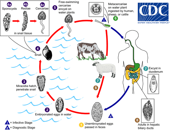
Adult liver flukes of the genus Fasciola have a characteristic leaf shape with an anterior oral sucker and a ventral sucker, and are found in the liver of ruminants and humans. The miracidium stage of the parasite looks nothing like the adult, because it is free living, covered with cilia and is "infectious" to snails. The cercaria even has a tail! But looks nothing like the adult or the miracidium. However, there is no denying that they are all the same species (Thomas, A.P., 1883). Suppose you sample water from a lake and find a miracidium, and suppose your atlas has no picture of a Fasciola miracidium, is it appropriate for you to name this parasite after yourself (which you can't technically do under the ICZN rules), and tweet #ifoundanewparasite?

Suppose alternatively that you have actually identified the miracidium as belonging to a trematode. How sure can you be that it is a miracidium of Fasciola and not the miracidium of Clonorchis or Dicrocoelium or Paragonimus? Are dichotomous keys based on overlapping length ranges enough for the identification?

The answer is no. You cannot definitively identify a miracidium without resorting to molecular techniques. 

Life cycle of Fasciola
The lifecycle of Fasciola, which also shows the morphology of life cycle parasites. Source: CDC
For more pretty pictures of the lifecycle, see a recent article in the Korean Journal of Parasitology @ http://parasitol.kr/journal/view.php?number=1870 


 And then there is the case of sexual dimorphism. For example, female oxyurids are larger than male oxyurids (Morand, 1998). If you were to find only a juvenile male as a result of your collection efforts, would it be right to compare its length to that of a female adult and declare the discovery of a new species or even a genotype, not realizing that your specimen is in fact a male?
And the alternate scenario, in which you identify it as a male, but have no explanation for why it is shorter than adult males are supposed to be.

The existence of species that look alike, even if their behavior is different poses another challenge to morphological identification. In the case of pathogenic parasites like Trypanosoma, if a veterinarian relies solely on morphology to distinguish between pathogenic T.brucei brucei (which causes nagana in cattle), and non pathogenic T.theileri, he is doomed, and so is the animal that he is attempting to treat, as is its owner. You may argue that the vectors for the two parasites are different. Yes. That is true. But the last time I checked, Trypanosomes on bovine clinical blood smears don't wear name badges that say which intermediate host they like to use.

The most logical thing is for us to ask ourselves, and each other these questions: 
  • How much should we rely on morphology to identify a rare parasite?
  • If a parasite has been described only once before, in a foreign language, should the claim be made that your new specimen is a new species, because of slightly different morphometrics? 
  • Is it correct to claim that you have identified a species using morphology, if you cannot produce photographs and reasons for the identification?
  • Is it okay to use a non-comprehensive atlas published in 1980 for the purpose of field identification?
  • Is it proper to not use microscopes in the field, because they are too heavy to lug around? 
  •  And since you are deep freezing your samples instantly after collection, it is perfectly acceptable to identify the parasites using morphology, a month after your return from field work. Right?
The correct response to the above questions is obviously "Not entirely" (for the first question) and "No" (for the other five). I hope those were your responses as well.  Supporting claims are mandatory when you are trying to identify rare parasites. It is perfectly alright to use morphology for identifying common parasites in clinical situations, solely on the basis of shape and features. Nobody would dispute the id of a large, pink-white, three lipped worm from the intestines of a pig as Ascaris, but doubts would arise if you found the same sort of worm in a fish and called that Ascaris

So, how now shall we id? The next time you tell someone that you like to identify rare parasites of an equally rare wild host species <insert your favorite host species here>, using morphometerics, it behooves you to back up your claims with supporting molecular data.
It is incumbent upon you to understand your data before you share it.

 
As always, opinions expressed are my own, you may choose to share them or choose not to.
 
References : 

Morand S., Hugot J.(1998) Sexual size dimorphism in parasitic oxyurid nematodes, Biological Journal of the Linnean Society 64,3,397-410
Thomas, A.P. (1883) The life history of the liver-fluke (Fasciola hepatica).QuarterlyJournal of Microscopical Science 23, 99–133


So, you made it to the end of the post! Great! As a reward, here is the picture of the type - specimen that is found in the alien's book.

From the best seller "Atlas of sentient life forms", from the publishing house Messier83, year Magellanic 0.197M, a collaborative effort of the Redshift consortium, page Q1W2E3 

Veterinarius parasitologistius - adult. Identification: Anterior part of body (known as "head" in the parlance of the organism) covered with numerous strands of black keratin, attached to which is a thin blue sparkling band. Two thin fore limbs seen, along with two hindlimbs which are capped in white, using which the organism is attached to the surface of the planet and which it uses for motility. The organism has a white outer layer, under which is found a blue layer. We are yet to ascertain which of these layers is the actual cuticle. The organism is uncommon, and is only found on the third planet orbiting the star Sol, in the Orion Arm of the spiral galaxy Milky Way.

PS: The picture is a representative of my lab attire.

Sunday, June 14, 2015

"Apicomplexans are lovely, dark and deep, but I have promises to keep, and miles to go before I sleep"

Dear Reader,
There has been a broadening of my research focus. 'Apicomplexity' has been a great blog to maintain, because that group of parasites is utterly fascinating. But, on the inside, I am a veterinarian through and through. Restricting myself to one group of parasites was not hard when I was working with a member of that group of parasites, that is, when I was working with Cryptosporidium, it was easy to restrict my blog to the Apicomplexans. But, a lot has happened since I first started this blog. I got a degree, moved places to start a PhD and have since begun working with other groups of parasites. And I definitely want to read, assimilate and write about the non-Apicomplexan parasitic beauties out there.

The point of this post is to inform you that you will be seeing more in this blog besides the Apicomplexans, including , but not restricted to ticks, and worms.

A new blog post is on its way !


Sunday, March 29, 2015

"The only good host is a live one", says Toxoplasma gondii

Immune evasion is the strategy that smart pathogens use to escape from the immune system and maximize their persistence and transmission. As has been alluded to, Toxoplasma is a perfect, intelligent parasite. It dampens the immune response, but allows just enough of an immune response to keep the host alive.


http://upload.wikimedia.org/wikipedia/commons/9/97/Toxoplasma_gondii.jpg
Credit: Image by Ke Hu and John Murray. Image is licensed under the Creative Commons Attribution 2.5 Generic license

The life cycle, briefly, of Toxoplasma gondii is as follows (in case you missed the myriads of posts that talk about Toxoplasma). Infective cysts when ingested, rupture in the new host, releasing the parasite. These invade the host cells and become the quickly replicating tachyzoite stage that cause the acute phase of the infection. This attracts the immune system, so that much of the infection is cleared. Some of the tachyzoites, however, persist, hitch rides to immunologically safe locations and become the slow replicating bradyzoites. These then form the bradycysts in important tissues like the CNS, the retina, the heart, causing a persistent chronic infection. When there is immunosuppression, the bradyzoites revert back and wreck havoc.

In an older article in Nature Reviews, the mechanisms of immune evasion and the recruitment of host anti-inflammatory pathways by the parasite are reviewed.

The author approaches the parasite from an evolutionary angle. Although the approach seems to give too much credit to a single celled protozoa, the anthropomorphic notion works well for the crafty Toxoplasma, the sole goal of which is to transmit itself to new hosts. Hence, it must do all that it can to multiply in its hosts, without killing them. To promote host survival, a powerful immune response is induced, which ensures that the host is not killed no matter what. But, Toxoplasma being clever, subverts the immune response. Thus, the very mechanism which ensures host survival is used to increase parasite persistence and transmission.

 The immune responses to Toxoplasma are summarized breifly below.

In the initial acute phase of the infection, NK cells are important for the initial immune response. IL12, which is secreted by the macrophages, neutrophils and DCs,  triggers the NK cells, and also triggers the interferon gamma dependent responses. DCs secrete IL12 when they harbor replicating Toxoplasma, or if they detect parasite derived molecules, even when other costimulatory signals are absent. Parasite derived molecules include cyclophilin 18 (a prolysylisomerase), which binds to the chemokine recptor CCR5 on DCs and trigger IL12 production. Incidentally, CCR5 also happens to be a coreceptor for HIV invasion, suggesting the hypothesis that C18 could inhibit monocyte infection by certain HIV strains.

TLRs take up some responsibility for IL12 induction. Evidence for this is provided by the fact that in MyD88 deficient mice, there is a marked reduction in the amount of IL12 secreted upon infection.TLR2 seems to  play a role in the overall immunity against Toxoplasma, with TLR2 deficient mice showing inefficient nitric oxide production by macrophages.MAP kinase p38 is involved in IL12 signalling. A Interferon inducible transcription factor (IRF8) promotes IL12 gene transcription. Absence of IRF8 also causes an absence of the DC subset CD8alpha +, which is  mainly involved in IL12 secretion.

So, why this intense fascination with IL12? What  exactly does IL12 do ?
IL12 activates NK cell and causes them to produce INF gamma, which in turn causes the proliferation of Type I CD4+ and CD8+ T cells, both of which also produce IFN gamma.

Macrophages infected with T. gondii produce nitric oxide in response to IFN gamma. Again, this does not completely eliminate the parasite, as some still escape exposure to NO.

Two strategies for immune evasion are mentioned, followed by an elaborate discussion of the second. The first is that the parasite becomes less susceptible to microbicidal activity by producing immunosuppressive molecules such as peroxiredoxins, that act against the effector molecules of the host. The second is that T. gondii uses mechanisms that exploit host anti-inflamatory responses.

Every effector mechanism in the body has a counteracting mechanism that keeps the former in check, ensuring homeostasis. This is true of the inflammatory response as well. A check to the wildly raging IFN gamma-dependent response is offered by IL10, which normally keeps in check the stimulated leukocytes in the acute phase of the infection. Now, IL10 is an antiinflammatory molecule that inactivates the microbicidal pathways triggered by IFN gamma, inhibits antigen processing and presentation by APCs, and inhibits cytotoxic cytokines producd by the T cells.

There is an upregulation of IL10 production that occurs concurrent to a T. gondii infection. If IL10 is neutralized in chronic Toxoplasmosis, severe CNS pathology results. Also, mice that produce little or no IL10 have uncontrolled INF gamma and TNF production in response to T. gondii, that results in severe leukocyte infiltration and hence severe inflammation. These mice also die in the acute phase of the infection. However, experts in the field disagree about the actual role played by IL10.

The second mechanism that is utilized is that of the potent antiinflammatory molecule lipoxin A4. LXA4 is generated by the action of 5-lipooxygenase on arachidonic acid via the intermediate leukotriene A4, which is acted upon by 15-lipoxygenase. Macrophages, but not DCs, participate in the production of lipoxins, even though the DCs are the main targets of LXA4. This mechanism operates independently of IL10. When mice that were IL10-deficient and those that were 5-lipoxygenase deficient, were challenged with T. gondii, severe infiltration with lymphocytes and severe necrosis of the liver without any CNS pathology was observed in the former, while there was infiltration of both the CNS and the liver in the latter. In mice that lacked 5-lipooxgenase, administration of IL 10 caused parasite reactivation and proliferation, because macrophage activity was inhibited.

The cellular source of 15-lipoxygenase, the enzyme that acts on leukotriene A4 to convert it to LX4, has been a mystery, suggesting that perhaps Toxoplasma itself produces the molecule. Proteomic analysis has showed that Toxoplasma tachyzoites carry a lipoxgenase that is similar to a plant derived type I lipoxygenase. Whether this is 15 lipoxygenase is yet to be confirmed. Also, the genes that code for the enzyme are yet to be identified in Toxoplasma. It is speculated that host cell invasion or immune attack of infected cells might trigger an upregulation of the lipoxygenase in intracellular forms.

5-lipoxygenase, the enzyme that converts arachidonic acid to leukotriene A4, is present in the host cells, and can be stimulated by parasite extracts. Since lipoxins dampen the immune response, host survival is ensured.

Two other examples of lipoxygenase modulation, that the author mentions, are: a pathogen coded lipoxygenases (a 15-lipoxygenase-like molecule) in Pseudomonas aeruginosa and a putative one in Mycobacterium tuberculosis.

In P. aeruginosa infections in cystic fibrosis patients, the authors speculate that the bacteria might use the lipooxygenase to produce lipoxins that might suppress inflammation, and so persist by not activating the immune system in immunocompetent patients. The actual relevance of this in clinical situations is yet to be elucidated because CF patients do not produce lipoxins in the lungs. The authors further speculate that mutations in the CFTR and other genes might also affect lipooxgenase generation directly in CF patients.

Mice that did not produce LXA4 endogenously seemed more resistant to infection with Mycobacterium tuberculosis.  They survived longer, had lower bacterial counts and a more pronounced type I CMI to the bacteria.

The author suggests that a contradiction emerges. Lipoxins seem to be protective in Toxoplama infections and yet detrimental in M. tuberculosis infections, suggesting that biological reasons are responsible. For Toxoplasma gondii, host survival is necessary to enable predation of the host. So, lipoxins dampen a fierce immune response, disallowing complete elimination of the parasite. In tuberculosis however, high bacterial numbers need to be generated to ensure transmission . Here, lipoxins dampen the immune response to allow proliferation.  The author concludes that lipoxin mediated immunomodulation is a new field with many unanswered questions and potential therapeutic applications.

Reference :
Aliberti, J., 2005. Host persistence: exploitation of anti-inflammatory pathways by Toxoplasma gondii. Nat Rev Immunol 5, 162-170.