Saturday, March 14, 2020

Let none say again that Dwarf pond snails are grasping and ungracious: Galba truncatula and Pseudosuccinea columella (Lymnaeidae)

A key feature of the life-cycle of trematodes of veterinary and medical importance is the use of snails as the first intermediate host. Typically, one learns the names of the various snails and goes on one's merry way. I thought it would be an intellectually engaging exercise to learn a little bit more about these intermediate hosts par excellence.

File:Galba truncatula shell.png
Galba truncatula 
By Peter Glöer & Vladimir Pešić
[CC BY 3.0  (https://creativecommons.
org/licenses/by/3.0)],
via Wikimedia Commons
Pseudosuccinea columella
By Francisco Welter Schultes ,
Public Domain,
https://commons.wikimedia
.org/w/index.php?curid=14726645
The intermediate hosts for the famous ruminant trematodes Fasciola and various Paramphistomes is an air breathing freshwater pond snail belonging to the genera Galba (previously Lymnaea) in the Family Lymnaeidae. Galba truncatula, commonly known as the dwarf pond snailis found in North and South America, Africa and Asia, and are notably absent in Australia. In the United States and Australia, Fasciola is transmitted by another snail called Pseudosuccinea columella. Another snail called Fossaria bulimoides can also host Fasciola. The vectorial capacity of Pseudosuccinea columella is marked because it can also be the first intermediate host of Heterobilharzia americana, the etiological agent of canine schistosomiasis in North America.  

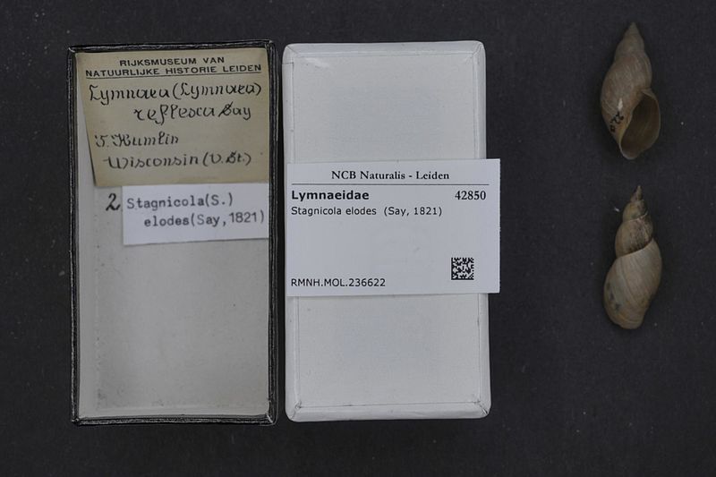
Like so many other organisms, the taxonomy of members of the family Lymnaeidae is under dispute, since Lymaeid snails are known to exhibit diversity and plasticity in shell appearance. Only 8 species inhabiting the Neotropics are considered as intermediate hosts of Fasciola hepatica, as described in a 2011 paper published in the journal Infection, Genetics and Evolution by Correa et alThese are Galba cousiniG. cubensisG. neotropicaG. truncatulaG. viatrixLymnaea diaphanaL. rupestris and Pseudosuccinea columella. It is also interesting to note that Lymnaea fuscus (Stagnicola fuscus) is partially resistant to infection with Fasciola hepatica while Stagnicola elodes is completely refractory.
Stagnicola fuscus which is partially
 resistant to infection by F. hepatica. 

Image attribution : Francisco Welter Schultes /
 Public domain via wikimedia.

File:Naturalis Biodiversity Center - RMNH.MOL.236622 - Stagnicola elodes (Say, 1821) - Lymnaeidae - Mollusc shell.jpeg
Stagnicola elodes which is completely
 refractory to infection with F. hepatica. 
Image attribution: Naturalis Biodiversity Center / CC0 via wikimedia

In general, Galba spp. can be identified by their turriform yellow to brownish shell of less than 7 - 10 mm height, an oval aperture less than 0.5 times the height of the shell, short spire, 5-6 inflated stepped whorls (the spiral structures of mollusc shells) and straight columellar edge. There may be striations on the shell.

Pseudosuccinea columella, also known as the American ribbed fluke snail, has a thin translucent shell that is brown with fine striations. It has 3.5-4 weakly convex whorls that end in a point. The aperture is oval with a straight columellar margin that is reflected only in the upper part. These snails may have white spots and measure 1.5 - 2 cm.

However, given that morphological identification beyond genus level may be hard, the use of molecular techniques is recommended. In the same paper, Correa et al. characterized the 18S, ITS-1, ITS-2 and CoxI genes of the 8 Neotropical species for ease of identification in cases where morphological characteristics are confusing/inadequate to identify a given lymnaeid specimen to species level.

So the next time you see a snail in stagnant waters/ water-associated environs such as reeds/mud, you can try to see if it is Galba or Pseudosuccinea.


References:

Title reference: The title of this post is a play on the common name of the snail (Dwarf pond snail) and a reference from the Lord of the Rings. Galadriel says " Let none say again that Dwarves are grasping and ungracious" to the parting fellowship (Ref here).


Correa AC, Escobar JS, Noya O, Velásquez LE, González-Ramírez C, Hurtrez-Boussès S, Pointier JP. Morphological and molecular characterization of Neotropic Lymnaeidae (Gastropoda: Lymnaeoidea), vectors of fasciolosis. Infect Genet Evol. 2011 Dec;11(8):1978-88. 

Animal base: http://www.animalbase.uni-goettingen.de/zooweb/servlet/AnimalBase/search


Friday, October 18, 2019

On improving our contributions to science

While several thousand primary research articles are published every year, secondary literature such as reviews are used to collect large amounts of research data into coherent, manageable pieces. They provide excellent sources of information for novices or for the seasoned veteran. Review writing is not just an art but a rigorous scientific task that must be systematically undertaken. In an article titled “On the benefits of systematic reviews for wildlife parasitology”, published in the journal International Journal of Parasitology Parasites and Wildlife in 2016, authors Haddway and Watson actively encourage authors of review articles to avoid non-systematic reviews.

They start by pointing out that systematic reviews require significant investments of time and resources to ensure that reliable methods are used to aggregate literature and understand the subject under study to avoid biases. In contrast, non-systematic reviews may exhibit biases including biases in publication selections, lowering their intended reliability. The authors provide an ideal example of how to conduct a systematic review in their paper. 

The best part of the paper is perhaps table 1 that lists the different types of reviews that they identified in wildlife parasitology, an aggregative field that combines ecology and parasitology.

1. “Configurative narrative integrative reviews” typically analyze qualitative metrics with little attention paid to quality and ill-defined criteria for inclusion or exclusion in the review. These are useful introductory notes to an area of work.

2. “Aggregative scoping reviews” typically analyze literature by vote-counting or categorizing but pay little attention to quality and possess ill-defined criteria for inclusion or exclusion. These provide a precursory evaluation of the scope of research in the field.

3. “Aggregative full literature reviews” typically are exhaustive searches that analyze quality data, placing them in categories based on some metric. These add to research in a field by synthesizing evidence provided in empirical data into coherent arguments.

4. “Aggregative meta-analysis reviews” typically use well-defined quantitative methods following exhaustive literature searches to synthesize statistically important arguments from multiple sets of empirical data, overcoming biases that arise from homogenous populations.

5.“Aggregative systematic reviews” are high in detail with quantitative and qualitative appraisals of all data available in a given area including grey literature, with assessments of quality and liability.

The authors expand on “domains” that affect the quality of reviews and give some advice for avoiding landmines that I think are best implemented during the writing process. I have summarized them here, but I suggest that the original paper be read for context (Link).

1. Transparency: To ensure that science remains repeatable and verifiable, the authors suggest that information on search strings and databases be provided.

2. Comprehensiveness: To avoid publication selection biases given the power of reviews in driving decision-making, the authors suggest that multiple databases be used with attention paid to non-significant results in grey literature.

3. Vote-counting: The authors posit that categorizing data into positive, negative or non-significant bins, aka vote-counting, should be avoided so that magnitude and variability of data that arises from heterogeneity and effect size are captured.

4. Critical-appraisal: Studies included in an review should be assessed for their reliability to ensure that only good quality data is included. The authors provide examples for why accuracy, validity and biological relevance may vary for each study based on its experimental design (sample size, control matching and inherent genetic variability).

5. Evidence for effects: The authors caution fellow writers on the over-arching tendency to declare the lack of evidence of effect as the evidence of no effect. They recommend extreme care in the interpretative phase so that a lack of studies is not interpreted as a lack of effect. 

I found that the advice prescribed in the article to be broadly applicable to review article writing in the biological sciences. It is especially relevant to graduate students writing their literature review chapters for their dissertations, and having recently written my own recently, I have found ways to improve it before final submission. Let us continue to make efforts to contribute to reliable science!

Reference:
Haddaway, Neal R., and Maggie J. Watson. "On the benefits of systematic reviews for wildlife parasitology." International Journal for Parasitology: Parasites and Wildlife 5.2 (2016): 184-191.


Sunday, March 17, 2019

A story of rotten apples and golden death!

What did a rotten apple in Paris, France and a rotten fig in Bangalore, India have in common?
They harbored common microscopic saprophytic worms that were host to the most remarkable worm-digesting bacteria.

 Biocontrol is the holy grail for environmentally conscious researchers. Biocontrol agents come in a wide variety of types, but the most common of them are predators, pathogens and parasitoids. Predators are of greatest use in pest control. Pathogens are very useful in weed control. Parasitoids have shown great success in lepidopteran insect control. 

Biological control has been endorsed by many as one of the ways to overcome resistance to anthelmintic drugs in livestock nematodes. Extensive research has been done with the microfungus Duddingtonia flagrans, which traps larvae in the pasture, and the bacterium Pasteuria penetrans, which has been used to control the root-knot nematodes of certain plants. The most recent event in this subfield of parasite control has been the isolation of a new bacteria called the golden death bacillus by a team of researchers based in Glasgow, Scotland. (Read the paper here).

Two isolates of the Golden death bacteria were isolated from worms found in a rotten apple obtained from Paris, France, and a rotten fig from Bangalore, India. The bacteria formed golden, mucoid colonies on agar and had a pungent odor. Microbiological testing and phylogenetic analysis of the whole genomes of the isolated bacteria revealed that they were very closely related but still distinct, and the name Chryseobacterium nematophagum was conferred on them, to describe both their color and their activity. 

The biological activity of Chryseobacterium nematophagum was experimentally studied in the model non-parasitic nematode C. elegans intially. C. elegans is a fantastic model for this study because it is a bacterivorous nematode that can be grown on bacterial lawns in the lab. Compared to control cultures of E. coli and a closely related isolate of Chryseobacterium gallinarum from a chicken, Chryseobacterium nematophagum was able to completely kill all L1 larvae placed on them within 7 hrs. As few as 200 bacteria were able to kill larvae in 24 hrs. C. elegans larvae were attracted to the bacteria in cultures, which is remarkable as they are repelled by other pathogens such as Serratia marcescens and Pseudomonas fluorescens. The larvae could not be killed by other related bacteria of the same family (Flavobacteriaceae). 

The authors observed that Chryseobacterium nematophagum multiplies in the pharynx of the C. elegans larvae and digested them from the inside out, which was demonstrated by the degradation of collagen and chitosan, but not beta-actin. The authors hypothesized that the effect of bacteria was due to chitinases and collagenases, and by comparative genomics of related bacteria were able to show that Chryseobacterium nematophagum indeed had copies of several nematode killing genes that encoded collagenases, chinases, cytolysins, hemolysins and others. Unique to Chryseobacterium nematophagum are slo- genes that code for a thiol activated cytolysins. Pertussis toxin S1 type secretion systems associated with motility and virulence were also found.

Chryseobacterium nematophagum was also able to kill bacterivorous, free-living larval stages of the important ruminant nematodes of the genera Haemonchus, Ostertagia, Cooperia and Trichoctrongylus, equine cyathostomins, and canine hookworm Ancylostoma among others. The bacteria, however, was not able to kill the potato nematode Globodera pallida or mosquito larvae.

Concerns on the safety of the bacteria to humans and animals have to be addressed before the bacteria can be deployed in full force in the field. We do not yet know if there are wild type Chryseobacterium nematophagum or similar bacteria already circulating in the pastures of the world, keeping nematode numbers in check. However, it is undeniable that the discovery of the golden death bacillus - Chryseobacterium nematophagum is a remarkable achievement.

References:  

Page, Antony P., et al. "The golden death bacillus Chryseobacterium nematophagum is a novel matrix digesting pathogen of nematodes." BMC biology 17.1 (2019): 10.

Stiling, Peter, and Tatiana Cornelissen. "What makes a successful biocontrol agent? A meta-analysis of biological control agent performance." Biological control 34.3 (2005): 236-246.

Sunday, February 3, 2019

Losses that help one develop : chromosomal diminution in ascarids

DNA losses are essential for the development of an organism. A familiar example of programmed DNA loss is the V-D-J joining in immunoglobulin genes in vertebrates that helps increase the repertoire of antibodies in mammals. In the ascarid nematodes, 'chromatin dimunition' is a form of programmed DNA loss that occurs during development at the embryonic 4 to 16 cell stage, eliminating large amounts of genomic DNA, but paradoxically adding to the total number of chromosomes. The phenomenon occurs only in five pre-somatic cells.

Although the phenomenon has been known since the 1880s, study of the fine details have only been possible in recent years after the advent of next generation sequencing. In a paper titled "Silencing of Germline-expressed genes by DNA elimination in Somatic Cells" published in the journal Developmental Cell (in 2012) , Wang et al. describe the phenomenon in Ascaris using a deep sequencing approach. The key take-aways from the paper are:
   
File:Chromosome en.svg
File:Chromosome-es.svg: KES47 (talk)
derivative work: KES47
[CC BY 3.0
(https://creativecommons.org/
licenses/by/3.0)],
via Wikimedia Commons
a. The sequence of events in chromatin diminution involve a break in the chromosome followed by loss of DNA and healing of the breaks by telomere addition to create new chromosomes.
  
b. In individual worms, the chromosomal break sites and DNA loss is pretty conserved even though there is a variation in the exact site of the break which occurs within 500 base pairs of each other in various tissues. Between individuals, this variation increases, with 70% of the breaks occurring with 1000 base pairs.
   
c. About 43 million base pairs are lost in total, with 12.7 million base pairs accounting for 685 unique genes expressed only during embryogenesis and/or gametogenesis.
   
d. Most of the eliminated genes play a role in mRNA translation and protein synthesis, and these jobs are taken up by similar "orthologous" genes in the larval and adult stages.
   
e. Chromatin diminution is not mediated by small RNAs like it is in ciliates, neither are there specific DNA elements in the chromosome that stipulate the break sites.

In a more recent paper titled "Comparative genome analysis of programmed DNA elimination in nematodes" published in the journal Genome Research (in 2017), Wang et al. divulge the secrets of chromatic diminution in nematodes closely related to Ascaris, namely Parascaris univalens and Toxocara canis. They show the following:

a. DNA elimination in Parascaris leads to a loss of 2.2 Gb (2.2 x 10^9 base pairs  of DNA, of which 10 Mb is unique.
  
b. In Toxocara canis,  approximately 49 Mb (49x10^6 base pairs) of DNA is eliminated, of which 20 Mb is unique.
   
c. The major repetitive sequences that are lost are a 120 base pairs satellite repeat in Ascaris, two short repeats of a 5 base pair sequence and a 10 base pair sequence in Parascaris and a 32-bp repeat in Toxocara canis.
   
d. The unique sequences that are lost include 1000 genes each in Ascaris and Parascaris, and 2000 genes in Toxocara canis. The increase in predicted genes lost in Ascaris over the previous study is of note. Since these genes are mainly expressed in the embryo, it is thought that this mechanism helps silence germline genes in nematodes.
    
e. The study also revealed that 86% of Ascaris genes and 88% of Parascaris genes occupy similar positions on the same chromosomes, which is also known as synteny. They also note that a set of 2623 genes are conserved between Ascaris, Parascaris univalens and Toxocara canis.
  
f. Chromosome break regions (CBRs) are the same in all individuals and occur at the same time in all five prismatic cells. These are regions of high SNP and indel densities. The breaks create new chromosomes, and the ends are "healed" by addition of telomeric ends. The authors identified 40, 46 and 32 CBRs in Ascaris, Parascaris and Toxocara respectively.
    
g. The authors were able to match 28 Ascaris CBRs to 26 CBRs in Parascaris, which is explained by synteny of genes. Such similarity did not exist in Toxocara.
     
h. The authors also investigated DNA accessibility in CBRs. They found no evidence of epigenetic changes but speculate that the reduced compactness of nucleosomes may lead to more accessible chromatin.
    
i. The authors provide preliminary evidence for changes in gene expression levels that occur as a result of proximity to telomeric ends. In Ascaris, they found that while expression levels of most genes in close proximity to the new telomeres do not change, some genes are silenced.
      
Both these papers describe an excellent application of the deep sequencing approach to the study of parasite biology. Chromatin diminution in nematodes, as far as we know, is an extraordinarily fascinating, precisely regulated occurrence in ascarid worms only. And as is usually the case, the more we know, the more we realize just how much we don't know yet.

References: [These are both on PMC. Click on the url to read the full articles]
Wang, J., Mitreva, M., Berriman, M., Thorne, A., Magrini, V., Koutsovoulos, G., Kumar, S., Blaxter, M. L., … Davis, R. E. (2012). Silencing of germline-expressed genes by DNA elimination in somatic cells. Developmental cell23(5), 1072-80. (https://www.ncbi.nlm.nih.gov/pmc/articles/PMC3620533/)

Wang, J., Gao, S., Mostovoy, Y., Kang, Y., Zagoskin, M., Sun, Y., Zhang, B., White, L. K., Easton, A., Nutman, T. B., Kwok, P. Y., Hu, S., Nielsen, M. K., … Davis, R. E. (2017). Comparative genome analysis of programmed DNA elimination in nematodes. Genome research27(12), 2001-2014. (https://www.ncbi.nlm.nih.gov/pmc/articles/PMC5741062/)

Friday, January 11, 2019

Spirocerca vulpis: New kid on the Spiruid block

In this genomic era when researchers are forever hunting for cryptic and hidden species, which differ ever so slightly from a well-established species, it is an extremely rare for someone to describe a new nematode from a common terrestrial mammalian host. Rojas et al. have done just that in a new article published in the journal 'Parasitology', and have successfully described and named Spirocerca vulpis from the red fox (Vulpes vulpes). Similar worms from the stomachs of red foxes have been reported in the past by Diakou et al. in Greece and Al-sabi et al. in Denmark, but had not been elevated to species status before.

When veterinary students are taught about the spirurids that reside in the GI tract, they are taught about a fascinating creature called Spirocerca lupi, an inhabitant of the oesophagus of canines and felines. Spirocerca lupi causes granulomas in the oesophagus and scarring of the aorta. The esophageal granulomas can become neoplastic and transform into fibrosarcomas and osteosarcomas. Migrations of the S. lupi away from the esophagus into other organs of the body can also occur rarely.

The new worm described by Rojas et al. differs morphologically from Spirocerca lupi . It is also located in a different predilection site - the stomach, while Spirocerca lupi is found in the esophagus. The authors report that a significant proportion (22%) of the fox population of Spain harbor smooth surfaced gastric nodules caused by Spirocerca vulpis, with one fox harboring a nodule in the pericardium. Spirocerca vulpis causes granulomas in the serosa or the wall of the stomach that grossly are grey-brown, smooth surfaced and surrounded by a zone of hyperemia. The adult worms live inside these nodules and are surrounded by an exudate. Worms of both sexes are reported to have six anteriorly projecting teeth and the females have embryonated eggs that resemble those of S. lupi. The oesophagus, which is a diagnostic feature in nematodes, is described as being cylindrical and muscular anteriorly and being glandular posteriorly. Analysis of the mitochondrial cox1 gene of the genus Spirocerca and closely related Cylicospirura and Protospirura revealed that S. vulpis was significantly different from S. lupi and the others. 

In addition, Rojas et al. in another paper titled "Phylogenetic analysis of Spirocerca lupi and Spirocerca vulpis reveal high genetic diversity and intra-individual variation" show that each individual worm in the genus Spirocerca has high individual variation in the nuclear ITS-1 locus, which is a compelling finding. It is well known that the internal transcribed spacers are multi-copy genes, with Ascaris possessing upto 40 copies per individual cell. It is also known that intra-individual variability exists in the ITS1 genes of Ascaris, as shown by Leles et al. in 2010. While Ascaris has 2-4 genotypes per individual, Spirocerca has unto 6 different ITS1 genotypes per individual.

Every new species that is described needs to be submitted to a collection for archival purposes. The type specimens for Spirocerca vulpis may be found in the National Natural History Collection of the Hebrew University of Jerusalem in Israel. It is now a valid species and you might find it in print as Spirocerca vulpis sp. nov., with 'sp. nov.' indicating that is a 'species novae' or a new species in the existing genera Spirocerca.

Some things to be gleaned from the papers are that (1) the species appears to be host restricted to red foxes (Vulpes vulpes), but it is unknown if other carnivores can be affected as no cross-transmission studies have been done yet. (2) It has also not yet been found/reported in North America, but an absence of a report is not always an indication of absence of existence in parasitology. (3) Both species of the genus Spirocerca are not known to be zoonotic. (4) Diagnosticians must add S. vulpis to their differentials list if they find a Spirocerca egg in red fox fecal samples. 

References:

Rojas, Alicia, et al. "Spirocerca vulpis sp. nov.(Spiruridae: Spirocercidae): description of a new nematode species of the red fox, Vulpes vulpes (Carnivora: Canidae)." Parasitology(2018): 1-12.

Diakou, Anastasia, et al. "First report of Spirocerca lupi infection in red fox Vulpes vulpes in Greece." Wildlife biology18.3 (2012): 333-336.

Al-Sabi, Mohammad Nafi Solaiman, et al. "Genetically distinct isolates of Spirocerca sp. from a naturally infected red fox (Vulpes vulpes) from Denmark." Veterinary parasitology 205.1-2 (2014): 389-396.

Rojas, Alicia, et al. "Phylogenetic analysis of Spirocerca lupi and Spirocerca vulpis reveal high genetic diversity and intra-individual variation." Parasites & vectors 11.1 (2018): 639.

Leles, Daniela, et al. "ITS1 intra-individual variability of Ascaris isolates from Brazil." Parasitology international 59.1 (2010): 93-96.


Thursday, December 13, 2018

Notes on the inclusion of Tetratrichomonas gallinarum in differential diagnosis lists

Granulomas as a disease process are used by some host organisms to wall off infectious agents, and may occur as a result of persistence of inflammatory stimuli for extended periods of time. In poultry, granulomas in visceral organs can be caused by a number of agents - Mycobacterium avium (which causes avian tuberculosis), granulomatogenic mucoid E. coli (which can cause coligranulomas), Yersinia pestis (pseudotuberculosis), Eubacterium tortuosum, Aspergillus fumigatus, and the parasites Histomonas meleagridis, larval migrations of the nematode Ascaridia, and by the cestode Raillietina echinobothridia. Another etiological agent that cause granulomatous reactions in poultry that often mimics the ones described above is Tetratrichomonas gallinarum.

In a guest editorial in the journal ‘Avian Pathology’ titled ‘Coligranulomatosis (Hjarre and Wramby’s disease) reconsidered’, authors Landman and van Eck give several reasons for why the diagnosis of Hjarre’s disease/coligranulomatosis in large outbreaks of granulomatous disease in poultry flocks is often actually a misdiagnosis (Ref 1). They posit that the parasitic protozoa Tetratrichomonas gallinarum must be considered on the differential list in such cases. Some of the reasons they list include:

a.  Coligranulomas generally occur sporadically in individual adult birds, with low flock prevalence, and are generally not of economic significance for poultry farming. 

b. Koch's postulates were never fulfilled for Hjarre's disease. The authors discuss several research studies that have successfully reproduced coligranulomatosis by infecting birds by injections of the infectious agent (by artificial routes - intravenous, intramuscular) but never by the natural oral route.

c. Several reports that the authors deem as misdiagnoses have highly variable mortality rates with several causing economic losses in the flocks, but none of these studies were able to reproduce coligranulomatosis experimentally by orally inoculating E.coli isolates from diseased birds  [and most were able to rule out tuberculosis as the cause]. 

d. The authors also cite reports of granulomatous disease in the caecum and liver in North American poultry flocks in which the etiology was described as unknown, because Koch postulates could not be fulfilled. These outbreaks had lesions consistent with a 2013 outbreak of granulomatous disease in poultry flocks in the Netherlands in which Landman et al. (Ref 2) fulfilled Koch’s postulates to demonstrate that the agent that caused upto 11% mortality in layers between 21 and 35 weeks of age was Tetratrichomonas gallinarum and not Escherichia coli

In their impressive study, Landman et al. (Ref 2) started with an investigation of seven affected layer flocks that showed a high incidence of mortality which were all linked to one rearing farm. They isolated and cultured the protozoa from affected birds, and identified it by morphology and PCR, and also use FISH probes to rule out bacterial and fungal agents. They undeniably proved the infectious nature of the agent by using sentinel hens on the affected farms that subsequently developed the same lesions as in the original outbreak. The authors fulfil another one of Koch’s postulates by experimentally inoculating material from the outbreak into specific pathogen free Leghorns intracloacally or intravenously, re-isolating the protozoa and confirming protozoan identity by PCR. 

The importance of both coligranulomatosis and Tetratrichomonas gallinarum mainly stems from the resemblance of the granulomas they produce to avian tuberculosis. Tetratrichomonas gallinarum as the sole causative agent has been subject to controversies because of subclinical infections, co-infections with Histomonas and age-related pathology due to which birds are affected only in the egg producing period. However, the paper and editorial by Landman et al. show that Tetratrichomonas gallinarum must be included in the differential diagnosis list in future outbreaks of graunulomatous disease alongside other well-known bacterial culprits. 


References:
1. W. J. M. Landman & J. H. H. van Eck (2017) Coligranulomatosis (Hjärre and Wramby’s disease) reconsidered, Avian Pathology, 46:3, 237-241, DOI: 10.1080/03079457.2017.1291903 

2. W.J.M. Landman, R.J. Molenaar, A. Cian, H.M.J.F. van der Heijden & E. Viscogliosi (2016) Granuloma disease in flocks of productive layers caused by Tetratrichomonas gallinarum , Avian Pathology, 45:4, 465-477, DOI: 10.1080/03079457.2016.1163325